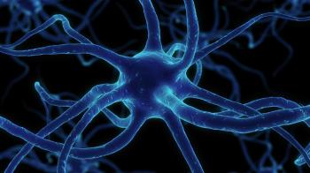
Merkezi Sinir Sistemi ve Kök Hücre Naklinde Nörit Büyüme İnhibitörlerinin Rolü
Zihinsel engelliliğe veya tam bir felce yol açan merkezi sinir sistemine zarar veren feci bir kaza, bazılarımız için ölümden daha kötü bir kaderdir. İnsanlar basitçe omuriliğini ya da beynini yenileyemeyeceği için bunun bir çaresi yok değil mi, yoksa var mı? Nöronlarını yenileyebilen bir organizma düşünecek olsaydık, aklımıza gelen ilk şey muhtemelen suda yaşayan semenderler olurdu. Ancak kendimizden başka bir yere bakmamıza gerek olmaması şaşırtıcı.
Periferik sinir sistemimiz, bir periferik sinir lifinin aksonu hasar gördüğünde, somada hasar olmadığı sürece kendini yenileme yeteneğine sahiptir. Bir periferik sinir lifinin aksonu parçalara ayrılırsa, soma ile bağlantısı olmayan kısımlar, bir zamanlar innerve ettikleri dokuya kadar dejenere olur, bu olaya Wallerian dejenerasyonu denir. Soma ile bağlantısı olmayan kısım dejenere olurken, soma ile bağlantısı olan kısım da retrograd dejenere denilen dejenerasyondan payını aldığı için dokunulmamış diyemeyiz. Aksonal hasarda sinir hücresinde görülen ilk değişiklikler somada olur ve buna kromatoliz denir, rejenerasyon ilerledikçe soma şişer ve çekirdek periferlere kayar. Wallerian dejenerasyonundan sonra, rejenerasyonun sonraki aşamaları, oligodendrositlerle yer değiştirdikleri için merkezi sinir sisteminde görülmeyen Schwann hücrelerinin proliferasyonu ile belirlenir. Santral sinir sisteminde nöronal aksonlar dejenere olduğunda aksonal rejenerasyon olmaz, gliosis olur. Periferik sinir sisteminin Schwan hücre kılıfı, merkezi sinir sistemine nakledildiğinde, nöronal aksonların tıpkı periferik sinir sisteminde olduğu gibi yenilenebildiği görüldü. Öyleyse, başından beri ana failimiz oligodendrositler miydi? Öyle olsa bile, nöral kök hücreler neden aşırı miktarda glial hücre oluşturmak yerine fonksiyonel sinir hücrelerine farklılaşmasın?
Nöroloji ve kök hücreler konusundaki bilgilerimiz son yıllarda giderek artsa da, nöral kök hücrelerin neye dönüşeceklerine dair seçimlerini belirleyen mekanizma henüz tam olarak aydınlatılamadı. Bu yazıda, merkezi sinir sistemindeki akson büyüme inhibitörlerine ilişkin mevcut anlayışımızın merkezini oluşturacak kadar önemli olan bir konuya değineceğiz: Nogo-A.
Nogo-A, MSS'nin önemli bir akson büyüme inhibitörüdür ve esas olarak oligodentrositlerde ifade edilir, ancak aynı zamanda büyük miktarda veriye göre Nogo-A’nın sadece oligodendrositlerle sınırlı olmadığını, aynı zamanda spinal motor nöronları, medulla spinalis dorsal kök ganglionları, sempatik nöronlar, retina ganglion hücreleri, neokorteks, hipokampus, purkinje nöronları, fibroblastlar, makrofajlar, miyoblastlar tarafından da ifade edildiğini biliyoruz. Nogo-A, retikülon 4 (RTN4, NOGO olarak da bilinir) geninin bir ürünüdür ve bu gen sadece Nogo-A'nın üretiminden değil, aynı zamanda diğer iki izoformu Nogo-B ve Nogo-C'nin de üretiminden sorumludur. Nogo-A ve bahsettiğimiz izoformları adlarından da anlaşılabileceği gibi MSS'nin nörit büyüme inhibitörleridir (Neurite outgrowth inhibitor). Nogo-A, endoplazmik retikulumda ve ayrıca miyelin kılıfının yüzeyinde de lokalizedir. Nogo-A nörit akson rejenerasyonunu engelleyen önemli bir alana sahiptir: Nogo-66. Nogo-66, hücre dışı yüzeyde ifade edilir ve iki hidrofobik alan arasında yer alan hücre dışı bir ilmektir. Nogo-66, etkisini göstermek için retikülon 4 reseptörü olarak da adlandırılan Nogo reseptörüne (NgR) bağlanır. NgR, ‘lösinden zengin tekrar proteinleri’ ailesine ait olan bir glikosilfosfatidilinositol (GPI) bağlantılı proteindir. P75NTR (nörotropin reseptörü p75), LINGO-1 (lösinden zengin tekrar ve immünoglobin benzeri alan içeren protein 1) içeren üçlü bir reseptör kompleksi oluşturarak sinyali iletir. Nogo-A'nın Nogo-66 alanının sinir aksonlarında NgR'ye bağlanması, büyüme konisinin yıkılmasına neden olur. Omurilik yaralanmalarından sonra aksonal rejenerasyonun inhibisyonu, NgR'nin tek etkisi değildir; bununla birlikte, aynı zamanda fizyolojik bir role de hizmet eder. Tavuk embriyolarının gelişimi esnasında komissural aksonların yol bulabilmesi için hem NgR1 hem de NgR3 gereklidir. NgR1 veya NgR3 kaybının aksonların orta hat bölgesinde durmasına neden olduğu ve bunun tavuk embriyosunda omurilik gelişimi esnasında çaprazlama sonrası aksonların rostral dönüşünü bozduğu bulunmuştur. NgR'lerin gelişimde fizyolojik etkileri olmasına rağmen, hayatımızın sonraki aşamalarında omurilik yaralanması olduğunda bizden yana olmadığı apaçık ortada, bu nedenle omurilik rejenerasyonunu yürütmek için bu reseptörü antagonize etme girişimleri vardır (IN-1 kullanımı gibi). Antikor tedavisine yönelik stratejiler şu anda geliştirilme aşamasındadır ve bu amaç için bir aşı geliştirmeye yönelik çalışmalar da bulunmaktadır. Ayrıca, rejenerasyonu teşvik etmek için nörotrofinlerin uygulanmasıyla periferik sinir parçaları veya schwann hücre köprüleri kullanılarak embriyonik kök hücrelerin veya daha matür hücrelerin nakledilmesi için yürütülen çalışmalar da vardır. Bu alandaki araştırmalar mevcut halleriyle çok umut verici olsalar da, transplante edilen kök hücrelerin nörolojik fonksiyonu geri kazandırabilmeleri için hem başarılı bir şekilde farklılaşmalarının hem de farklılaşırken operasyonel bağlantılar kurmalarının gerekmesi ve farklılaşmamış kök hücrelerin transplantasyon sonrası kontrolsüz bırakıldıklarında teratokarsinom oluşturma eğilimlerinin olması gibi birçok sorunla karşı karşıya kalmaktadırlar.
~~~
Neurite Outgrowth Inhibitors’ Role at Stem Cell Transplantation in Central Nervous System
A catastrophic accident causing damage to the central nervous system leading to a mental disability or a complete paralysis is a fate worse than death for some of us to consider. As humans can’t just simply regenerate their brain or spinal cord there is no remedy to this, or is there? If we were to think of an organism capable of regenerating its neurons the first thing that would come to our minds would probably be the aquatic salamanders. But much to our surprise we don’t need to look any further than ourselves.
Our peripheral nervous system is capable of regenerating when a peripheral nerve fiber’s axon gets damaged as long as the soma is scatheless. If a peripheral nerve fiber’s axon gets cut in pieces the parts that have no connection to the soma becomes degenerated all the way up to the tissue they were once innervating, this phenomenon is called Wallerian degeneration. While the part that had no connection to the soma gets degenerated, the part that has connection to the soma is not untouched either since it also gets its share of degeneration called retrograde degeneration. In axonal damage the first changes seen on the nerve cell happens at the soma and its called chromatolysis, as the regeneration progresses the soma becomes swelled and the nucleus slides to the peripheries. After Wallerian degeneration the later stages of regeneration are determined by Schwann cells proliferation, which is not seen on the central nervous system as they are replaced by oligodendrocytes. When neuronal axons degenerate at central nervous system axonal regeneration does not happen but the gliosis happens. When the Schwan cell sheath of the peripheral nervous system was transplanted into the central nervous system the neuronal axons were capable of regenerating just like they could on the peripheral nervous system. So was the oligodendrocytes were our main perpetrator all the way along? Even if then, why wouldn’t the neural stem cells differentiate into functional nerve cells rather than creating an excess of glial cells?
Although our knowledge in neurology and stem cells has increased exponentially in the recent years the underlying mechanism of the neural stem cell’s fate decision is yet to be fully elucidated. In this article we will touch to an important subject matter that’s so important that it is the central of our current understanding of axon growth inhibitors in central neural system: Nogo-A.
Nogo-A is an major axon growth inhibitor of the CNS and its mainly expressed in oligodentrocytes yet we also know a large amount of data has shown that Nogo-A is not limited to the oligodendrocytes but its also expressed in spinal motor neurons, dorsal root ganglion, sympathetic neurons, retinal ganglion cells, neocortex, hippocampus and purkinje neurons, fibroblasts, macrophages, myoblasts. Nogo-A is a product of reticulon 4 (RTN4 also known as NOGO) gene and it is not only responsible of the production of NogoA but also the other two isoforms Nogo-B and Nogo-C all of which are hence the name neurite outgrowth inhibitors of CNS. Nogo-A is localized in endoplasmic reticulum and also in the surface of myelin sheath, It contains an important domain that inhibits neurite axon regeneration : Nogo-66. Nogo-66 is expressed at the extracellular surface and it is a extracellular loop located between the two hydrophobic domains. In order to exert its effects Nogo-66 binds to Nogo receptor (NgR) which is also named as reticulon 4 receptor. NgR is a glycosylphosphatidylinositol (GPI) anchored protein that belongs to the family of leucine-rich repeat proteins. It transduces signal by forming a tripartite receptor complex that contains P75NTR (neurotrophin receptor p75), LINGO-1 (leucine-rich repeat and immunoglobin-like domaincontaining protein 1). The binding of the Nogo-A’s Nogo-66 domain to the NgR at the nerve axons induces growth cone collapse. The inhibition of axonal regeneration after spinal cord injuries is not the only contribution of the NgR; however, it also serves a physiological role. Both NgR1 and NgR3 are required for commissural axon pathfinding during development. It was found that loss of NgR1 or NgR3 causes axons to stall in the midline area and to interfere with the rostral turn of postcrossing axons during spinal cord development of the chicken embryo. Although NgRs have their physiological effects at the development they certainly don’t serve well when there is spinal cord injury at the later stages of our life so there are attempts at antagonizing this receptor (such as with the use of IN-1) to conduct spinal cord regeneration. Strategies for delivering antibody treatment are currently under development and additionally there are studies for developing a vaccine for this cause. There are also studies conducted for transplanting embryonic stem cells or more mature cells by the use of fragments of peripheral nerves or schwann cell bridges with the administration of neurotrophins to promote regeneration. Although the researches in this area are very promising, in their current state they face many issues such as the need to form operational connections while successfully differentiating in order to restore neurological function or the undifferentiated stem cell’s tendency to form teratocarcinomas when left not regulated after transplantation.
Yazan: Umut KÖLENİŞ
Kaynakça
Chen, M. S., Huber, A. B., van der Haar, M. E., Frank, M., Schnell, L., Spillmann, A. A., ... & Schwab, M.
E. (2000). Nogo-A is a myelin-associated neurite outgrowth inhibitor and an antigen for monoclonal antibody IN-1. Nature, 403(6768), 434-439.
Wang, B., Xiao, Z., Chen, B., Han, J., Gao, Y., Zhang, J., ... & Dai, J. (2008). Nogo-66 promotes the differentiation of neural progenitors into astroglial lineage cells through mTOR-STAT3 pathway. PloS one, 3(3), e1856.